Harnessing Spin Coating to Create Aligned Hydroxyapatite Bone Scaffolds
Reproducing the complex, layered structure of natural bone in the lab has long challenged materials scientists. At University College London (UCL), Dr. Jishizhan Chen, working with Professors Wenhui Song and Jia Hua, set out to emulate bone’s “Bouligand” architecture—where collagen and hydroxyapatite (HAp) crystals are arranged in concentric, oriented layers—to guide stem-cell behavior and drive uniform mineral growth. In this post, we’ll explore how Navson Technologies’ precision spin coater enabled this breakthrough, the cellular responses it provokes, and what it means for next-generation bone implants.
The Challenge of Bone Anisotropy
Natural cortical bone achieves its remarkable combination of strength and toughness through a highly ordered, layered structure of collagen fibrils interspersed with HAp nanocrystals. Most synthetic scaffolds, by contrast, suffer from randomly oriented minerals that compromise mechanical fidelity and directional strength.
Navson Spin Coater: Precision Alignment at Scale
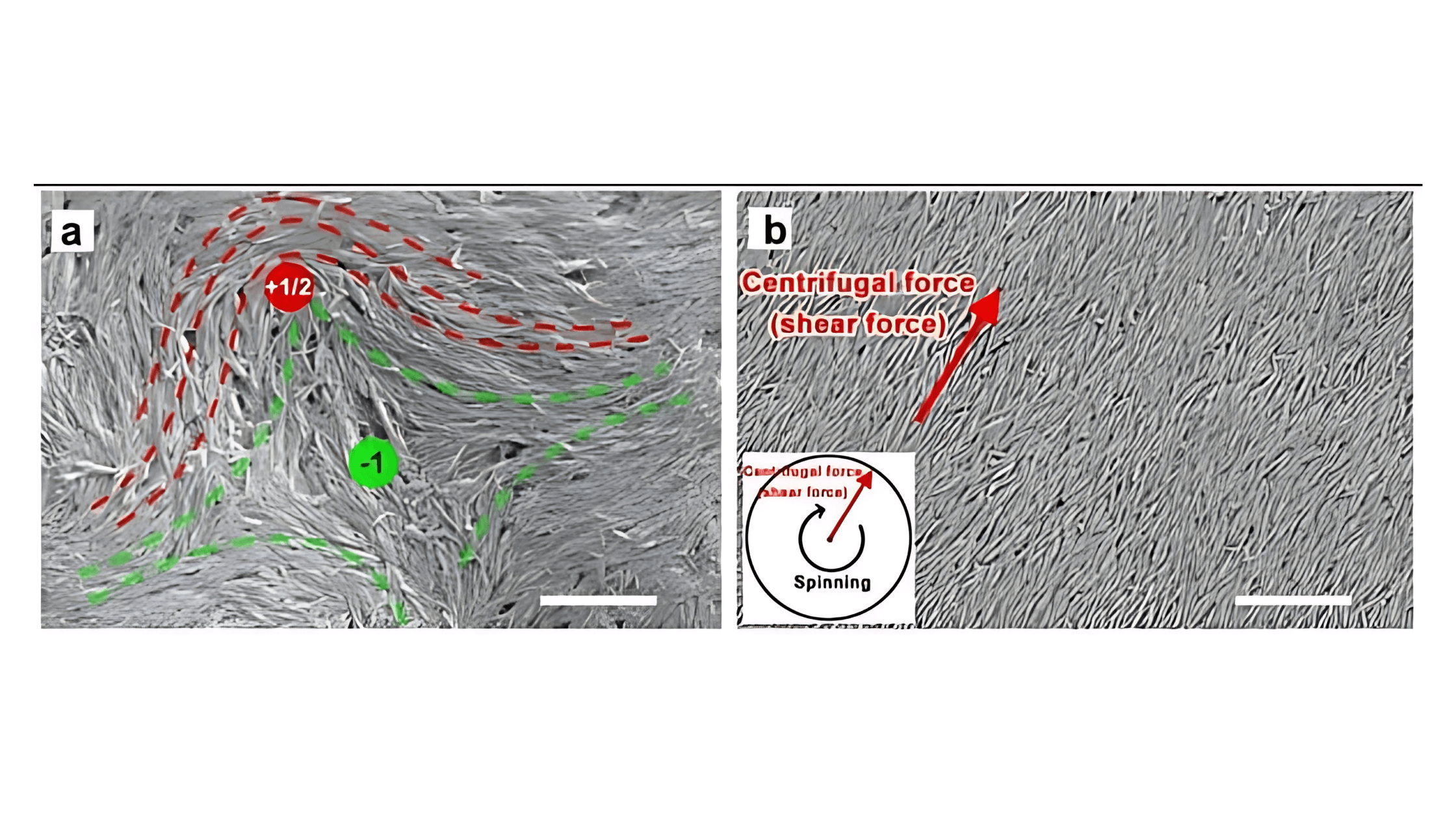
Using Navson’s NT12000 spin coater, Dr. Chen deposited uni-directional, liquid-crystalline films of citrate- and terbium-doped HAp nanorods. By fine-tuning spin speed and solution viscosity, he achieved centimeter-scale films where every nanorod aligns parallel to the centrifugal force—replicating bone’s natural (002) crystal orientation in seconds rather than hours.
Why spin coating?
- Centrifugal shear forces align rod-shaped nanoparticles rapidly
- Delivers large-area uniform films
- Compatible with standard lab workflows for scalable substrate fabrication
Directing Stem-Cell Fate with Nanoscale Topography
On these aligned HAp substrates, human bone marrow stem cells (hBMSCs) exhibited:
- Morphological alignment along the rod axis within days
- Upregulation of ECM genes (COL1A1 & COL4A6) to organize collagen networks
- Activation of the PI3K-Akt pathway, boosting osteogenic markers (ALP, OCN, RUNX2)
Compared to random controls, uni-directional films produced 25–40 % more calcium deposition by day 28—forming the first ordered nanolamellar calcium sheets (≈36 nm spacing) templated by the aligned ECM and HAp rods.


Molecular Mechanisms of Ordered Mineralization
RNA-seq and bioinformatics mapping revealed how topography translates into biology:
- Focal adhesion sensing: Aligned rods concentrate integrin binding
- Signal transduction: COL1A1/COL4A6 → F-actin tension → PI3K-Akt cascade
- Secondary templating: Oriented collagen guides stacked calcium nanosheets into multi-layer bone-like assemblies.